An endotracheal tube, commonly referred to as an ETT or tracheal tube, is a thin, flexible plastic tube that is inserted through the mouth or nose down into the trachea (windpipe). Its main purpose is to maintain an open airway and allow a patient to breathe when they are unable to do so unaided. This article will delve deeper into what endotracheal tubes are used for, how they work, and some important aspects about their use and care.
Uses of Endotracheal Tubes
Endotracheal Intubation, which involves inserting an ETT, is often required during surgery when a patient needs to be put under general anesthesia. This is because under anesthesia the muscles of the throat relax and can collapse, obstructing the airway. Therefore, securing an artificial airway with an ETT is necessary to allow breathing while the person is unconscious.
ETTs are also commonly used in emergency situations when a person is unable to maintain their own airway, such as during cardiac arrest, major trauma, severe breathing difficulties, or drug overdoses. By establishing a clear pathway into the lungs, the ETT helps provide oxygen and allows ventilation by bag-mask or mechanical ventilator.
For patients with respiratory conditions such as COVID-19, ETTs may be necessary to apply Positive End-Expiratory Pressure (PEEP) therapy or to connect the patient to an Extracorporeal Membrane Oxygenation (ECMO) machine, both of which can help improve oxygen levels in the blood. ETTs are also useful for laryngoscopy procedures that require examination or treatment within the vocal cords and trachea.
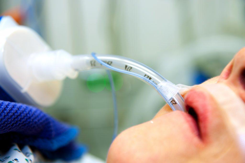
Components and Placement of Endotracheal Tubes
An ETT typically consists of a safely flexible tube made of polyvinyl chloride (PVC) plastic, with an inflatable cuff or balloon at its distal tip. The proximal end has a standard 15mm connector to attach to a ventilator circuit, anesthesia machine or bag-mask resuscitator.
To insert an ETT, the vocal cords are visualized using a laryngoscope, which holds the tongue out of the way. The tip of the lubricated ETT is then gently guided between the vocal cords and advanced downward a proper predetermined distance into the trachea. Correct placement in the mid-trachea is confirmed using methods like observation of condensation in the tube, chest rise with ventilation, auscultation of breath sounds and capnography monitoring of exhaled carbon dioxide.
Once situated, the cuff at the distal end is inflated with air using a syringe to form a tight seal within the trachea. This sealing function of the cuff is crucial, as it helps prevent air from leaking up and around the ETT instead of entering the lungs during ventilation. It also prevents stomach contents and secretions from entering the trachea and lungs.
Nursing Care of Endotracheal Tubes
Optimal care and maintenance of ETTs is important to promote patient safety and comfort. Some key nursing interventions involve:
- Securing the ETT with tape or a commercial securing device to prevent accidental dislodgment.
- Inflating the cuff with the minimum volume of air needed to form an effective seal and prevent excess pressure on tracheal walls.
- Suctioning secretions above and below the cuff as needed to keep the airway clear.
- Repositioning the patient as able and performing oral hygiene regularly to prevent pressure sores around the mouth.
- Monitoring cuff pressure using a manometer and inflating if needed to maintain an adequate seal without injury.
- Checking placement and ensuring proper ventilation and breath sounds periodically.
- Documenting details of ETT care like cuff pressure, positioning changes and suctioning thoroughly.
- Involving speech therapy as indicated to prevent laryngeal injury from intubation.
Potential Complications of Endotracheal Tubes
While ETTs are life-saving devices, their use does carry some risks if not carefully managed. Potential complications can include:
- Aspiration: If the cuff leaks or is not properly inflated, stomach contents can be inhaled into the lungs causing infection.
- Tracheal injury: Over-inflating the cuff can damage the tracheal mucosa, potentially leading to stenosis over time. Improper technique during intubation also poses trauma risks.
- Ventilator-associated pneumonia: Pneumonia risk increases the longer someone requires ventilation, as saliva and secretions can pool above the cuff.
- Accidental dislodgment: If accidental extubation occurs, the airway may need to be resecured emergently.
- Laryngeal injury: Prolonged intubation can cause laryngeal nerve damage, vocal cord injury or laryngeal edema on extubation.
Close monitoring and following best practices for ETT care help reduce risks. Overall, with proper usage, ETTs serve as an invaluable medical advancement for airway management in numerous critical care situations. Further research continuously improves their safety profile as well.
Endotracheal tubes are vital medical devices that help facilitate breathing and oxygenation in patients who cannot maintain their own airway. From intubation technique and tube positioning to ongoing nursing care and potential risks, this article aimed to provide thorough insight into endotracheal intubation procedures and management of ETTs. Their applications significantly benefit patients in emergencies, surgery and respiratory distress. With standardized best practices, ETTs save lives on a daily basis worldwide.
Get more insights on this topic: