Flow cytometry is a powerful tool that allows researchers to analyze individual cells in a fluid stream. This technique enables detection and characterization of cells according to their optical and fluoresence properties. Over the past few decades, flow cytometry has revolutionized research in immunology, hematology, and oncology
Working Principle
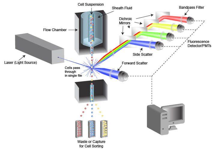
Fluidic system: Flow cytometry relies on using a special fluidic system to move cells in single file within a stream of fluid. Cells are suspended in a fluid and injected into the flow cytometer as a liquid suspension. A flow cell carries the stream of cells at speeds of several meters per second. This aligns cells spatially so they can pass one by one through the interrogation point.
Laser-based detection: As cells pass through the flow cell, they are intercepted by one or more lasers. Laser light scattered from each cell is collected by optical detectors. Forward scattered light provides information about cell size, while side scattered light relates to complexity or granularity of internal structures.
Fluorescence detection: Cells can be stained with fluorescent dyes or antibodies that have been conjugated to fluorescent molecules. Different fluorescent probes bind to specific markers on cells like proteins or DNA. When laser light excites these probes, they fluoresce at characteristic wavelengths. Multiple fluorescence detectors collect emitted light to detect different fluors simultaneously from each cell.
Data processing: Flow cytometers have fast digital electronics and computers attached to process immense data from thousands of cells analyzed per second. These computer systems record characteristics like scatter and fluorescence of each individual cell as it passes through the instrument. Data analysis software allows classification, quantitation, and characterization of cell populations.
Applications of Flow Cytometry
Cell cycle analysis and apoptosis: Flow cytometry is widely used to analyze cell cycle distribution and quantify apoptosis or programmed cell death. After proper staining, it enables detection of different phases of cell cycle based on DNA content. It also distinguishes between live, dead, early and late apoptotic cells.
Immunophenotyping: Cell surface and intracellular markers play important roles in immune cell characterization and disease diagnosis. Flow cytometry combines multiple fluorescent antibodies for immunophenotyping of leukocytes. This helps in subclassification of lymphocytes and their activation states. It is crucial for detection, monitoring and management of hematological malignancies.
Stem cell research: Stem cells have important applications in regenerative medicine. Flow cytometry is crucial for isolation of stem cell populations based on specific surface markers. It also allows purity assessment after in vitro expansion and differentiation protocols. Human embryonic and induced pluripotent stem cells are routinely characterized by flow.
Microparticle analysis: Microparticles are membrane vesicles released from activated or apoptotic cells. Their protein and RNA cargo provides clues about cellular processes. Flow cytometry enables sensitive quantification and phenotyping of microparticles from biological fluids in various disease states. This emerging area holds promise for novel diagnostics.
Research applications: Apart from clinical applications, flow cytometry finds extensive use in basic research. It allows detection of reporter gene expression, protein-protein interactions, calcium signaling and metabolic activity assays in cells. Combining flow with cell sorting enables purification of rare cell populations for ‘omics’ studies or to generate cell lines.
Latest Advances
In the last decade, flow cytometry technology has undergone tremendous advances. New generation flow cytometers offer more lasers, more fluorescence channels, higher sensitivity photodetectors, and faster throughput rates. Cell sorting speed and precision have also improved greatly. Computational advances facilitate analysis of high-dimensional datasets and discovery of novel cell populations. Development of microfluidic flow platforms, mass cytometry using metal isotopes as labels, and imaging flow cytometry are expanding the capabilities of this technique. With continual technological innovation, flow cytometry will remain a key workhorse for cell-based research and diagnostics.
flow cytometry has revolutionized cellular analysis due to its ability to measure multiple parameters from thousands of individual cells within short times. Starting from basic enumeration of immune cell subsets, its applications have grown manifold to include multiparametric analysis of cellular phenotypes, functions, signaling, and tracing of cellular dynamics. Advances in instrumentation and computational tools will further strengthen this versatile technique. With integration of artificial intelligence and microfluidics, flow cytometry promises to deliver deeper biological insights and enhance precision medicine approaches in the years ahead.
Get more insights on this topic: https://www.newsanalyticspro.com/flow-cytometry-a-powerful-tool-for-scientific-research-and-medical-diagnosis/