Diagnostic Tests
If a person exhibits potential symptoms of lung cancer, their doctor will recommend certain tests to determine if cancer is present and, if so, the extent or stage of the disease. Common diagnostic tests for lung cancer include:
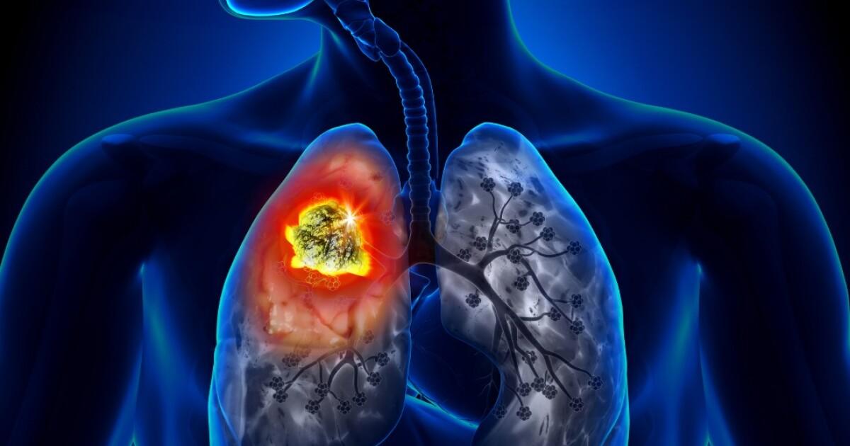
- Chest X-ray: An X-ray produces images of the structures in the chest, including the lungs, and can reveal abnormalities. However, chest X-rays alone cannot determine if a suspicious spot is cancerous.
- CT (computed tomography) scan: A low-dose CT scan takes detailed images of the lungs and surrounding structures. It is more sensitive than chest X-rays and better able to detect early-stage lung cancer as well as secondary tumors in other organs.
- MRI (magnetic resonance imaging) scan: An MRI uses magnetic fields and radio waves to produce cross-sectional Lung Cancer Diagnostic And Screening images of organs and tissues in the chest area. It is used in some cases to determine the stage of lung cancer.
- PET (positron emission tomography) scan: A PET scan provides information about whether cancer cells in the lung are actively growing. The person receives an injection of a small amount of radioactive substance (tracer) and the scan detects where the tracer accumulates. Regions of cancerous activity appear as bright spots on the scan.
- Sputum cytology: A sample of sputum, or mucus coughed up from the lungs, is examined under a microscope to check for cancer cells.
- Needle biopsies: If an abnormality is suspected based on imaging tests, the doctor may perform a needle biopsy to obtain tissue or cell samples for examination. Types include bronchoscopy, CT-guided needle biopsy, endobronchial ultrasound, and thoracentesis.
Sponsored
Lung Cancer Diagnostic and Screening: Overview
Sponsored